

 Akshay Dinesh
Fat is usually described as the body’s long-term energy store, but that description leaves out a small and unusually active tissue. Brown adipose tissue, better known as brown fat, spends energy to make heat. It is especially important in babies, who cannot rely on strong shivering to stay warm, and researchers now know that many adults retain working deposits of it around the neck, collarbones, spine, and major blood vessels.
Brown fat is not a magical calorie burner. Adults generally have far less of it than white fat, its activity varies widely, and much of what scientists hope it could do for human health remains under study. Yet it offers a striking example of how the same basic fuel can meet two very different needs. One fat cell saves energy for later; another can release that energy as warmth within moments.
A large study published in Nature in October 2026 has renewed interest in this tissue. The researchers used medical images and records from more than 100,000 people, then followed an unexpected drug association into cell and mouse experiments. The result is promising as a route for future research, but it also shows why evidence must be separated carefully from treatment claims.
White fat stores fuel; brown fat is built to spend it
Most body fat is white adipose tissue. A typical white fat cell holds one large droplet of triglyceride, a compact way to store more energy than the body needs immediately. Between meals or during exercise, enzymes can release fatty acids from that reserve. White fat also acts as an endocrine organ, sending chemical signals that help regulate appetite, inflammation, insulin sensitivity, and other processes.
Brown fat cells look and behave differently. They contain many smaller lipid droplets and an unusually high number of mitochondria, the cell structures that convert energy from food into forms cells can use. Iron-containing proteins inside those mitochondria give well-supplied brown tissue its darker colour. Dense networks of blood vessels deliver oxygen and fuel, then carry the newly produced heat into the circulation.
The tissue’s main job is non-shivering thermogenesis: heat production without repeated muscle contractions. Newborns have prominent brown-fat deposits because their small bodies lose heat quickly and their muscles cannot yet generate enough warmth through shivering. Adults usually retain smaller, patchier deposits, most often above the collarbones and deep in the neck. Some white-fat depots can also develop heat-producing cells called beige adipocytes when exposed to signals such as repeated cold.
These categories are useful, but real tissue is not arranged in three perfectly separate boxes. Brown and beige cells vary by location, age, genes, hormones, temperature history, and health. What unites the heat-producing cells is their ability to divert mitochondrial activity away from making as much usable chemical energy and toward releasing more of that energy as heat.
A mitochondrial shortcut turns a fuel gradient into warmth
Mitochondria normally use energy from nutrients to pump protons across an inner membrane. The resulting concentration difference works rather like water held behind a dam. When protons flow back through the enzyme ATP synthase, the cell captures that stored potential by producing ATP, the molecule that powers much of its work.
Brown-fat mitochondria contain large amounts of uncoupling protein 1, or UCP1. When UCP1 opens a path through the inner membrane, protons can return without driving ATP synthase. Fuel is still oxidized and oxygen is still consumed, but less of the released energy becomes ATP. More leaves as heat. Biologists call this uncoupling because fuel oxidation is partially disconnected from the usual energy-capturing step.
The process is controlled rather than permanently switched on. A brown fat cell must keep its own machinery running, so it cannot simply waste every gradient it builds. During cold exposure, nerve signals help release fatty acids from the cell’s small lipid droplets. Those fatty acids provide fuel and also help activate UCP1. Brown fat can draw on glucose and circulating fats as its activity rises, while its many capillaries distribute the heat.
That mechanism explains why describing brown fat as tissue that merely “burns calories” is incomplete. Its immediate biological purpose is temperature control, not weight loss. The energy cost is real, but the total effect in a person depends on how much active tissue is present, how strongly it is stimulated, how long it stays active, and whether the body compensates through hunger or other changes.
Cold starts a whole-body response, not a single-cell trick
Temperature sensors in the skin and body send information to the hypothalamus, a brain region that coordinates temperature regulation. If the body needs more heat, the sympathetic nervous system releases norepinephrine at nerve endings within brown fat. The signal reaches receptors on brown adipocytes, starts the breakdown of stored triglycerides, and increases the activity of the thermogenic machinery.
Brown fat works alongside other defences. Blood vessels near the skin narrow to reduce heat loss. Muscles may shiver, posture changes, and behaviour does much of the practical work: people add clothing, move indoors, or seek a warmer place. Non-shivering thermogenesis is one contributor to a coordinated response rather than an independent thermostat.
Repeated cool exposure can increase the activity of existing brown tissue and recruit more thermogenic features in some fat cells. That does not make deliberate cold exposure a risk-free health strategy. Responses differ greatly among people, and severe cold can lead to hypothermia, heart strain, frostbite, or other harm long before any speculative metabolic benefit becomes relevant. Research protocols use controlled temperatures, screening, and monitoring for a reason.
Brown-fat activity also follows patterns that complicate simple comparisons. It is often easier to detect in younger and leaner people and during colder seasons, but an association does not prove which factor caused another. Medications, hormones, sleep, diet, illness, and the temperature of the scanning room can all affect what researchers observe.
Medical scans overturned an old assumption about adults
For much of the twentieth century, brown fat was treated mainly as a feature of newborns and small mammals. Pathologists had seen brown-looking tissue in some adults, but its importance was uncertain. The decisive evidence arrived through a tool designed for a different purpose: positron emission tomography combined with computed tomography, or PET/CT.
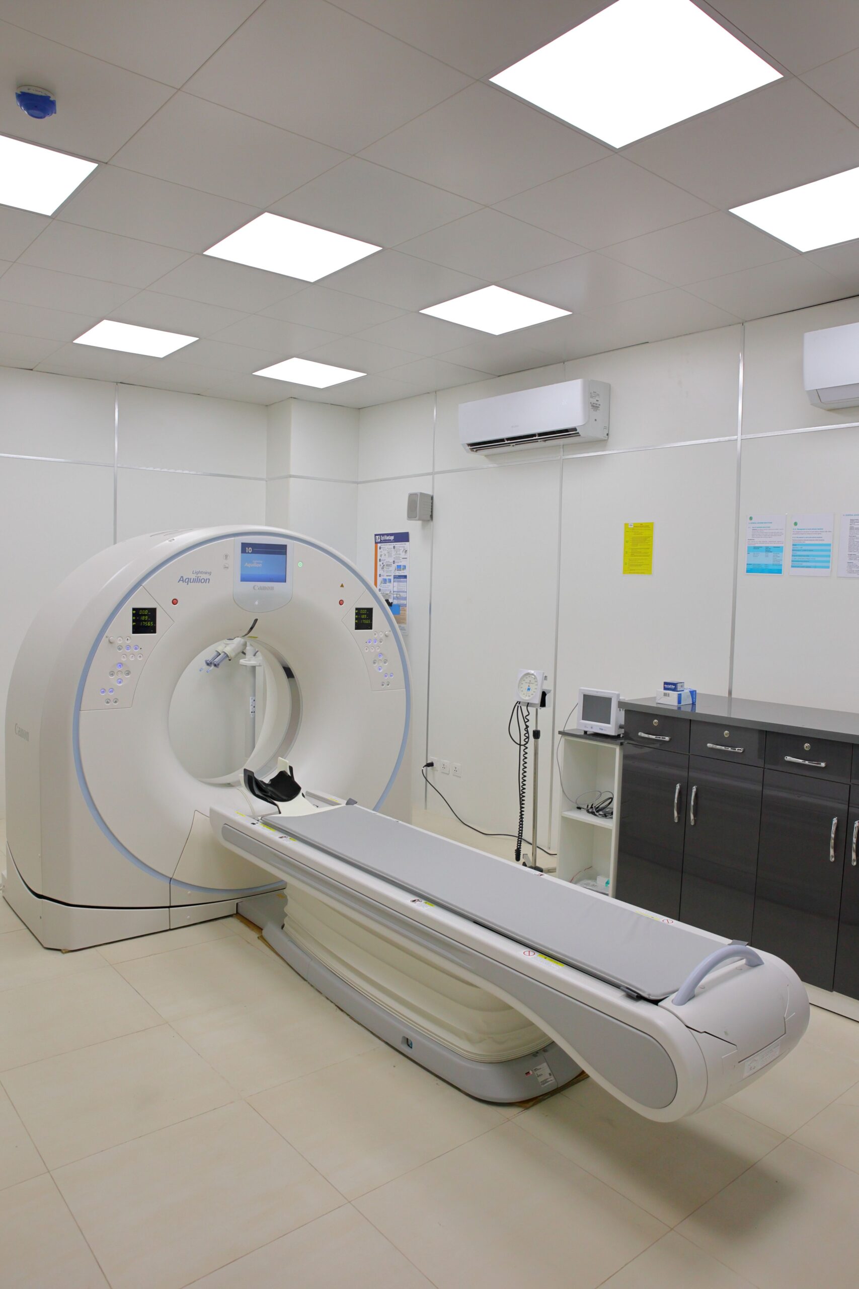
Many cancer scans use a radioactive glucose analogue called 18F-FDG. Tissues that take up a great deal of glucose appear as active regions on the PET image, while CT shows their anatomical location. Radiologists sometimes saw symmetrical bright patches above the collarbones that were not tumours or lymph nodes. The patches often appeared more strongly in cold weather and matched the density of fat on CT.
Several research groups reported in 2009 that these adult deposits contained active brown fat. Biopsy samples expressed UCP1, and controlled cooling made the tissue easier to detect. Those findings changed a question from “Do adults have brown fat?” to “How much do they have, how active is it, and what does it do beyond keeping them warm?”
PET/CT is powerful, but it does not provide a perfect census. A scan commonly tracks glucose uptake, while active brown fat can use several fuels. Preparation, medication, recent temperature, and the chosen detection threshold can influence the result. Cancer-centre records also come from patients who are not necessarily representative of the wider population. The images reveal valuable patterns, but their context matters.
What the new drug finding does, and does not, show
The 2026 study led by Nicolás Gómez-Banoy and Paul Cohen expanded the clinical view substantially. Their team analysed PET/CT images and medical records from 101,243 patients at a large cancer centre. A machine-learning method helped quantify brown-fat activity in about 8,700 of them rather than classifying tissue only as present or absent. Greater detected activity was associated with more favourable measures of glucose, blood lipids, inflammation, and heart function, as well as lower recorded prevalence of several cardiometabolic conditions.
The researchers also found that people taking angiotensin receptor blockers, a family of medicines used to treat high blood pressure, were more likely to show brown fat. Losartan stood out for further testing. In cultured brown adipocytes, it increased markers of thermogenic activity and oxygen consumption. Male mice given losartan showed higher energy expenditure during the experiment, including during a cold challenge, without a change in body weight.
Those steps make the finding more informative than a database correlation alone, but they do not turn it into a proven human therapy. The patient analysis was retrospective, so unmeasured differences between groups may have influenced the associations. The cohort came from a cancer centre. The direct activation experiments used cells and mice, and the mouse dose and conditions do not tell people how a prescribed human dose would affect brown fat. No clinical trial in the study showed that starting losartan activates brown fat or improves metabolism in people.
Losartan is a prescription medicine with established uses, dosing rules, interactions, and possible adverse effects. The research is not a reason to take it without a medical need or to change an existing treatment. Its scientific value lies elsewhere: it points researchers toward a molecular pathway that may be separable from blood-pressure lowering. That idea can now be tested prospectively and, if the evidence continues to hold, may guide the design of more targeted compounds.
Brown fat remains compelling because it links cell biology to an everyday experience: the body must decide whether to save chemical energy or release it as heat. UCP1 reveals how that decision works at the mitochondrial membrane, PET/CT shows where the active tissue persists in adults, and large clinical datasets can expose leads that smaller experiments might miss. The newest lead is worth following precisely because it is still a question. Good research turns an unexpected association into a testable mechanism, then asks human trials to determine whether that mechanism can become useful medicine.